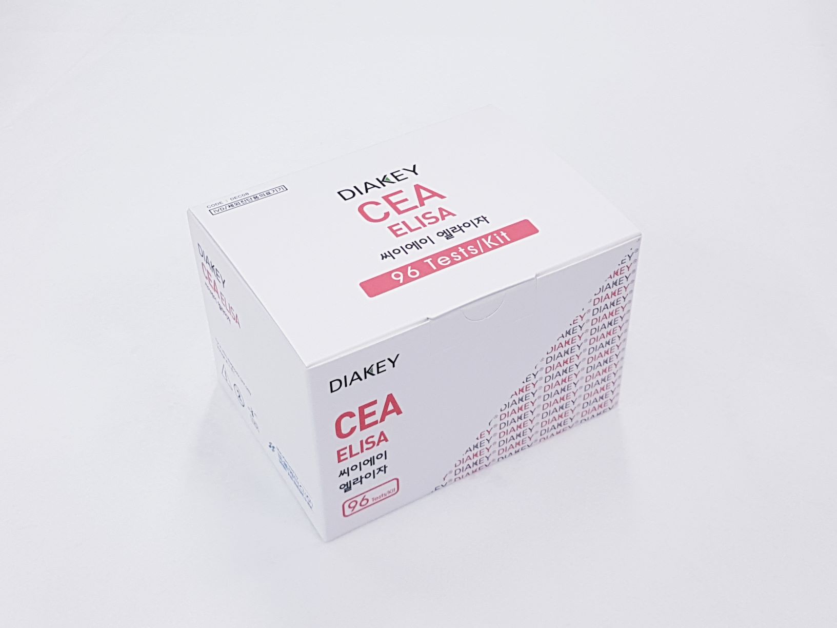
DIAKEY CEA ELISA
Enzyme immunometric assay for quantitative determination of carcinoembryonic antigen (CEA) in human serum or plasma
Summary
-
KFDA Registration No
14-3093
-
CAT No
DEC08
-
TEST METHOD
ELISA
-
SAMPLE VOLUME
50 ul
-
INCUBATION TIME
30'+10'RT
-
STD RANGE
0-100 ng/ml
Download File
Intended Use
Enzyme immunometric assay for quantitative determination of carcinoembryonic antigen (CEA) in human serum or plasma
INTRODUCTION
The carcinoembryonic antigen (CEA) is a glycoprotein soluble in perchloric acid, first identified by Gold and Freedman in embryonic endodermal tissues of 2-6 month old feti and in epithelial tumors of endodermal origin (gastrointestinal tract). CEA molecule presents a high degree of heterogeneity, depending on the carbohydrate contents (50-60%) and also influenced by the purification method. Heterogeneity results clear also from molecular weight (175,000-2,000,000 daltons), sedimentation coefficient (6.2-6.8 S) and isoelectric point (p.l. 3-4), while it’s electrophoretic mobility is always beta. CEA immunological determination allowed evidencing of CEA-like molecules, a family of molecules with common antigenic determinants, synthesized by normal and pathological tissues, among which the most relevant one is NCA (Non-specific cross reacting antigen). Use of monoclonal antibodies has made it possible to definitely overcome the problem of the cross-reacting CEA like molecules when assaying CEA.
PRINCIPLE OF THE ASSAY
The DIAKEY CEA ELISA is a solid-phase non-competitive immunoassay based upon the sandwich technique. Standards, controls, and samples are incubated together with a biotinylated and HRPO labeled Anti-CEA antibody in streptavidin coated micro stripes. CEA present in Standards or samples is adsorbed to the streptavidin coated micro strips by the biotinylated anti-CEA antibody during the incubation. Unbounded material is removed by a washing step. After washing, substrate reagent is added to each well and the enzyme reaction is allowed to proceed. During the enzyme reaction a blue color will develop if antigen is present. The intensity of the color is proportional to the amount of CEA present in the samples. The color intensity is determined in a microplate spectrophotometer at 450nm (reference 620nm). The CEA concentrations of samples are read from the calibration curve.
Use Precaution
Be careful when handling all samples, reagents, or devices used in the test as they may be the source of infection.
All reagents, human body samples, etc. are handled at the designated location.
- Do not use mixed reagents from different lots.
- Do not use mixed reagents from different lots.
- Use distilled water stored in clean container.
- Use an individual disposable tip for each sample and reagent, to prevent the possible cross-contamination among the samples.
- Rapidly dispense reagents during the assay, not to let wells dry out.
- Wear disposable globes while handling the kit reagents and wash hands thoroughly afterwards.
- Do not pipette by mouth.
- Do not smoke, eat or drink in areas where specimens or kit reagents are handle.
- Handle samples, reagents and loboratory equipments used for assy with extreme care, as they may potentially contain infectious agents.
- When samples or reagents happen to be spilt, wash carefully with a 1% sodium hypochlorite solution.
- Dispose of this cleaning liquid and also such used washing cloth or tissue paper with care, as they may also contain infectious agents.
- Avoid microbial contamination when the reagent vial be eventually opend or the contents be handled.
- Use only for IN VITRO.

